Aragen Bioscience offers a diverse range of preclinical oncology models and services with client-specific customized study design. These include human xenograft tumor models as well as the more complex sub-renal capsule, and syngeneic tumor models. We monitor PK/PD correlations using plasma/serum following test article administration, tumor response, changes in tumor volume and weight, as well as studying genomic, proteomic and metabolomic biomarkers.
Xenograft models are extensively used in investigational new drug (IND)-enabling studies for evaluation of new chemical entities (NCEs) and new biological entities (NBEs) as potential anticancer agents. Our scientists have developed and validated numerous xenograft models in Ncr Nu/Nu, NOD-SCID and SCID-Beige mice. We will also customize and utilize human cell lines of your interest to conduct in vivo proof of concept studies. Syngeneic models enable testing anticancer agents in an immunocompetent system, although they may not entirely reflect the human immune competency. With intact immune system, syngeneic models are pertinent for evaluating immunologically based targeted therapies alone or in combination.
We have expertise and facilities to support your pre-clinical oncology research with our extensive range of in vivo models enlisted below:
| Human Cell lines @ Aragen Bioscience (USA/India) | |||||
|---|---|---|---|---|---|
| S.No | Cell line | Origin of Tumor | Species | USA | IND |
| 1 | A431 | Epidermoid carcinoma | Human | Y | Y |
| 2 | Hep G2.2.15 | Hepatocellular carcinoma | Human | Y | |
| 3 | A-673, A-673-Fluc | Rhabdomyosarcoma (Ewing tumor) | Human | Y | |
| 4 | KASUMI-1 | Acute myeloid leukaemia | Human | Y | Y |
| 5 | MV4-11 | Beta myelomonocytic leukemia | Human | Y | |
| 6 | NALM-6 | B-Cell leukemia | Human | Y | |
| 7 | SUDHL-10 | B-Cell leukemia | Human | Y | |
| 8 | MDA-MB-468 | Breast cancer | Human | Y | |
| 9 | MDA-MB-231, MDA-MB-231-Fluc | Breast cancer | Human | Y | Y |
| 10 | MCF-7, MCF-7-Fluc | Breast cancer | Human | Y | |
| 11 | SK-Br-3 | Breast cancer | Human | Y | |
| 12 | Raji, Raji-Fluc-GFP | Burkitt’s lymphoma | Human | Y | |
| 13 | FaDu | Cervical carcinoma | Human | Y | |
| 14 | HT-29 | Colon adenocarcinoma | Human | Y | |
| 15 | HCT-116, HCT-116-Luc | Colon adenocarcinoma | Human | Y | |
| 16 | Colo 205 | Colon adenocarcinoma | Human | Y | |
| 17 | SW480 | Colon adenocarcinoma | Human | Y | |
| 18 | KM-12 | Colon adenocarcinoma | Human | Y | |
| 19 | MKN-1-Fluc | Gastric cancer | Human | Y | |
| 20 | U87-MG, U87-MG-Fluc | Glioblastoma | Human | Y | |
| 21 | Hep 3B | Hepatocellular carcinoma | Human | Y | |
| 22 | PLC/PRF/5 | Hepatoma | Human | Y | |
| 23 | A549, A549-Fluc-GFP | Lung cancer (NSCLC) | Human | Y | |
| 24 | NCI-H460 | Large cell lung carcinoma | Human | Y | |
| 25 | NCI-H226 | Lung cancer (NSCLC) | Human | Y | |
| 26 | H1299 | Lung cancer (NSCLC) | Human | Y | |
| 27 | NCI-H358 | Lung cancer (NSCLC) | Human | Y | |
| 28 | NCI-H1944 | Lung cancer (NSCLC) | Human | Y | |
| 29 | NCI-H1573 | Lung adenocarcinoma | Human | Y | |
| 30 | Calu-6 | Lung anaplastic carcinoma | Human | Y | |
| 31 | NCI-H292 | Mucoepidermoid pulmonary carcinoma | Human | Y | |
| 32 | A427 | Lung carcinoma | Human | Y | |
| 33 | A375 | Melanoma | Human | Y | |
| 34 | KG-1 | Multiple myeloma | Human | Y | |
| 35 | NCI-H929 | Multiple myeloma | Human | Y | |
| 36 | U266 | Multiple myeloma | Human | Y | |
| 37 | MM1.S | Multiple myeloma | Human | Y | |
| 38 | RPMI8226 | Multiple myeloma | Human | Y | |
| 39 | WSU-DLCL-2* | Non-Hodgkin’s lymphoma | Human | Y | |
| 40 | IGROV-1* | Ovarian cancer | Human | Y | Y |
| 41 | SK-OV-3, SK-OV-3-Fluc | Ovarian cancer | Human | Y | |
| 42 | A2780 | Ovarian cancer | Human | ||
| 43 | OVCAR-3 | Ovarian adenocarcinoma | Human | Y | Y |
| 44 | OV90 | Ovarian cancer | Human | Y | |
| 45 | PANC-01 | Pancreatic cancer | Human | Y | |
| 46 | AsPC-1 | Pancreatic cancer | Human | Y | |
| 47 | BxPC3, BxPC3-Fluc-GFP | Pancreatic cancer | Human | Y | |
| 48 | PANC.10.05 | Pancreatic cancer | Human | Y | |
| 49 | MIA PaCa-2, MIA-PaCa- 2-Rluc | Pancreatic cancer | Human | Y | |
| 50 | 22rv.1 | Prostate cancer | Human | Y | |
| 51 | LNCaP | Prostate cancer | Human | Y | |
| 52 | DU-145 | Prostate cancer | Human | Y | |
| 53 | PC-3 | Prostate cancer | Human | Y | Y |
| 54 | 786-O, 786-O-Fluc | Renal cell carcinoma | Human | Y | |
| 55 | A498 | Renal cell carcinoma | Human | Y | Y |
| 56 | Caki-1, Caki-1-Fluc | Renal cell carcinoma | Human | Y |
| Animal Cell lines @ Aragen Bioscience (USA/India) | |||||
|---|---|---|---|---|---|
| S.No | Murine Cell Line | Origin of Tumor | Species | USA | IND |
| 1 | 4T1, 4T1-Fluc | Breast cancer | Mouse | Y | |
| 2 | MC-38, MC-38-Fluc | Colon cancer | Mouse | Y | |
| 3 | CT-26, CT-26-Fluc | Colon cancer | Mouse | Y | |
| 4 | B16-F10 | Melanoma | Mouse | Y | |
| 5 | B16-F1 | Melanoma | Mouse | Y | |
| 6 | Hepa 1-6 | Hepatoma | Mouse | Y | |
| 7 | LL/2 | Lung cancer | Mouse | Y | |
| 8 | A20 | Lymphoma | Mouse | Y | |
| 9 | EL4 | Thymoma | Mouse | Y | |
| 10 | E.G7-OVA | Ova-expressed thymoma | Mouse | Y | |
| 11 | C6VL | T-cell lymphoma | Mouse | Y | |
| 12 | L1210 | Leukemia | Mouse | Y | |
| 13 | Renca | Renal cortical adenocarcinoma | Mouse | Y | |
| 14 | Ba/F3 | a murine interleukin-3 dependent pro-B cell line | Mouse | Y | |
| 15 | D17 | Osteosarcoma | Canine | Y |
| S.No | Cell Line | Origin of Tumor | Type of Model | Species | USA | IND |
|---|---|---|---|---|---|---|
| 1 | A549-FLuc-GFP | Lung cancer (NSCLC) | Lung metastasis | Human | Y | |
| 2 | BxPC3-FLuc-GFP | Pancreatic cancer | Orthotopic | Human | Y | |
| 3 | Mia Paca-2-RLuc | Pancreatic cancer | Orthotopic | Human | Y | |
| 4 | U87-MG-Luc | Glioblastoma | Orthotopic | Human | Y | |
| 5 | MDA-MB-231-FLuc | Breast cancer | Orthotopic | Human | Y | |
| 6 | SKOV-3-FLuc | Ovarian cancer | Orthotopic | Human | Y | |
| 7 | Raji-FLuc-GFP | Burkitt’s lymphoma | Disseminated xenograft | Human | Y | |
| 8 | 4T1-FLuc | Breast cancer | Lung metastasis | Mouse | Y | |
| 9 | GL-261-Luc | Glioblastoma | Orthotopic | Mouse | Y |
| S.No | Tissue Type | Human Cell Line |
|---|---|---|
| 1 | Leukemia lymphoma | Daudi, MV4.11 |
| 2 | Burkitt’s lymphoma | Raji, Raji-Luc-GFP |
| 3 | Acute myeloid leukemia | Kasumi-1, KG-1 |
| 4 | B-cell leukemia | NALM6, SUDHL-10 |
| 5 | Multiple myeloma | NCI-H929, U266, MM.1s, RPMI-8226 |
| 6 | Colon adenocarcinoma | HT-29, HCT-116, HCT-116 Luc-GFP, Colo 205, SW480 |
| 7 | Lung cancer (NSCLC) | A549, NCI-H226, NCI-H1299, NCI-H358, NCI-H1944, A549-Luc-GFP |
| 8 | Lung anaplastic carcinoma | Calu-6 |
| 9 | Lung adenocarcinoma | NCI-H1573, NCI-H292, NC-H460 |
| 10 | Renal cell carcinoma | Caki-1, 786-O |
| 11 | Hepatocellular carcinoma | Hep G2, Hep 3B |
| 12 | Pharynx squamous cell carcinoma | FaDu |
| 13 | Pancreatic cancer | BxPC-3, BxPC-3-Luc-GFP, MIA PaCa-2-Luc |
| 14 | Ovarian cancer | SKOV-3, SKOV-3-Luc, OVCAR-3 |
| 15 | Prostate cancer | LNCaP, LNCaP-Luc, 22rv.1 |
| 16 | Breast cancer | MDA-MB-468, MDA-MB-231, MDA-MB-231-Luc, MCF-7, MCF-7 Luc, BT-474 |
| 17 | Glioblastoma | U-87MG-Luc |
| 18 | Melanoma | A-375 |
Additional Models
- Lung metastasis model
- Orthotopic models of breast, pancreas, and brain
- Sub-renal capsule assay
- Vaccine model
Other Oncology Support
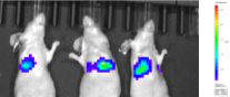
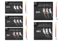
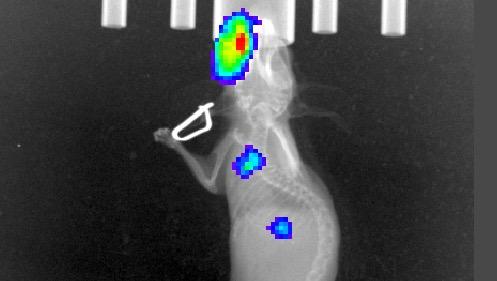
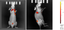
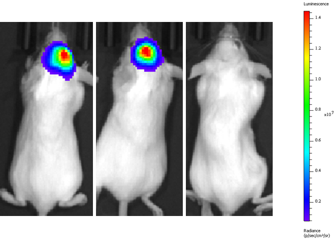
- In vivo bioluminescent imaging
- 70+ cancer cell lines of validated models stored in-house
- Pharmacokinetics and bioavailability of various test materials in tumor-bearing mice
- Target engagement of compound in tumor tissue and PK/PD correlation
- Maximum tolerated dose (MTD) studies in immunocompromised mice
Typical readouts are, but not limited to:
- Tumor growth kinetics
- Immunophenotyping with multicolor FACS
- Cytokine panels
- ELISpot assays
- Multi-gene analysis
- Histology/ immunohistochemistry
- Protein/ peptide analytics
These services cater to a range of therapeutic areas including immuno-oncology research.
Immuno-oncology (IO) and CAR T-Cell Therapeutics Evaluation Capabilities
In recent years, immuno-oncology research has made rapid progress, thanks to new therapeutic options such as CAR T-cell. While the CART therapy is increasingly being used for the treatment of human hematologic malignancies, there are several challenges including targeted delivery, immunogenicity, and efficacy in solid tumors.
To address these challenges and help you expedite your IO research projects to market, we have developed various mouse models to screen potential CAR T-cell therapeutics to support IND-enabling process.
Aragen Bioscience is a leading integrated discovery, development, and manufacturing solutions provider for the global life sciences industry. Our Discovery Biology Services include in vitro and in vivo screening of new immunotherapies, including engineered chimeric antigen receptor (CAR) T-cells using translational animal models to support efficacy studies in xenograft and syngeneic tumor models.
Validated Immuno-oncology Model
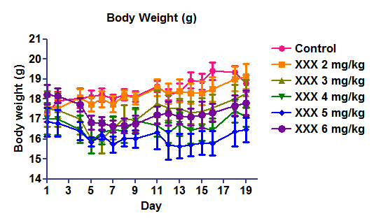
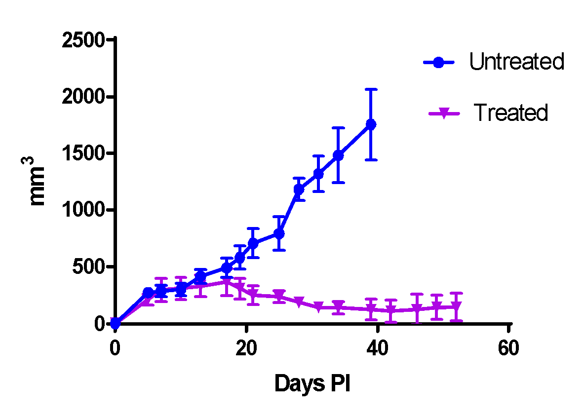
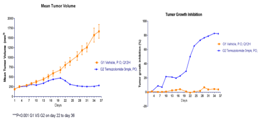
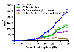
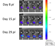
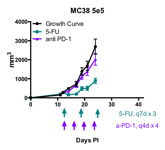
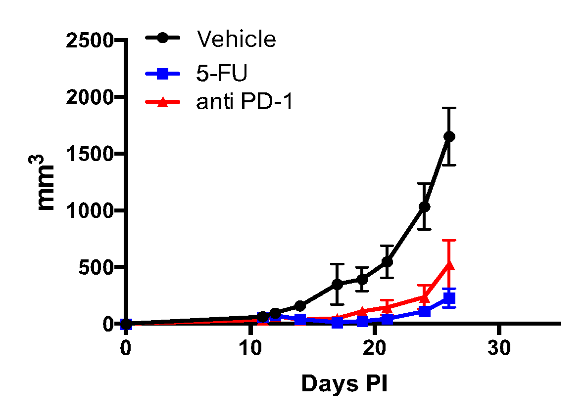
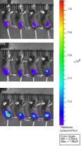
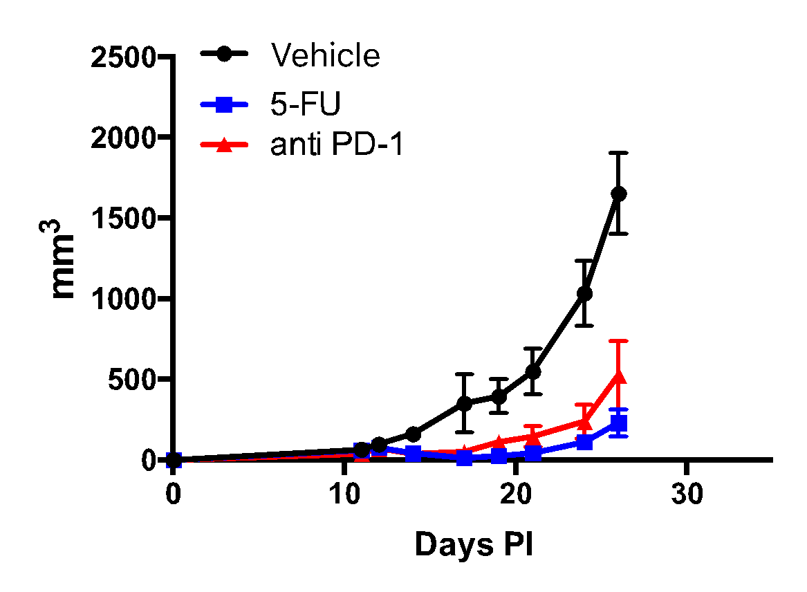
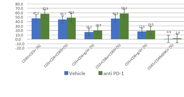
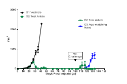
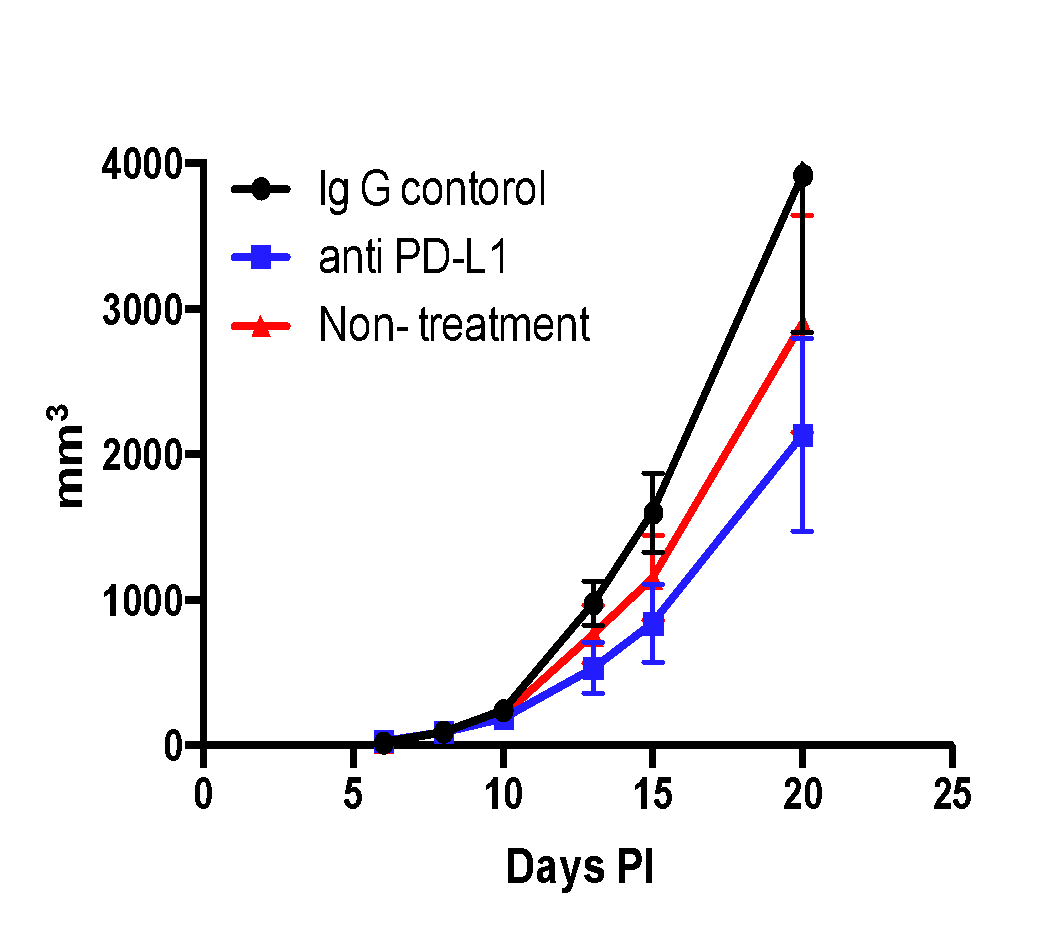
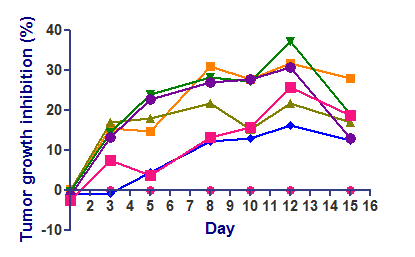
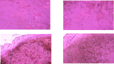
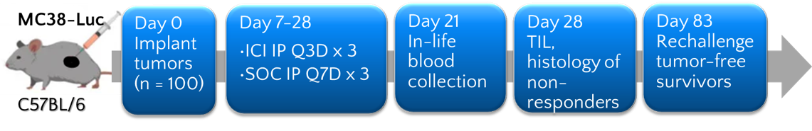
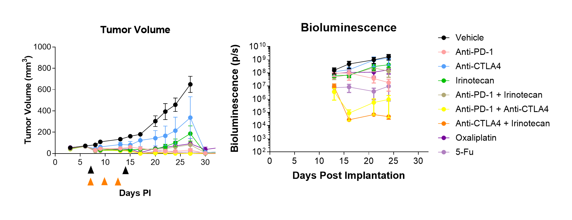
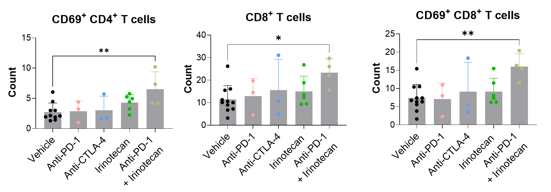
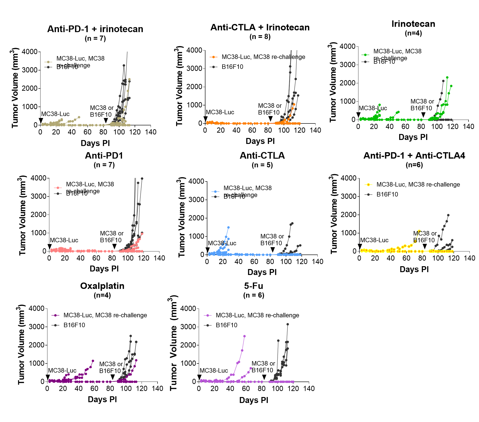
MC38 cells represent a widely recognized murine colon adenocarcinoma cell line extensively employed in preclinical investigations, notably within the realm of immuno-oncology. Using the luciferase-tagged MC38 that was developed at Aragen Bioscience, we successfully replicated the effects of anti-PD-1, anti-CTLA-4, and their combination in the subcutaneous MC38 tumor model. In addition, all three standard-of-care chemotherapeutics significantly reduced tumor volume compared to the vehicle group. For the first time, we demonstrated that anti-CTLA-4 alone and in combination with irinotecan prevented subcutaneous MC38 but not B16F10 tumor growth in re-challenged responders, suggesting a specific immunity toward MC38 tumor cells. Our study demonstrated that subcutaneous MC38- Luc tumor model is a robust model for testing multiple drug modalities including immunotherapy and chemotherapy.
The Aragen Bioscience Advantages
- Experienced and skilled team of in vivo research scientists to support pre-clinical research from study design through execution.
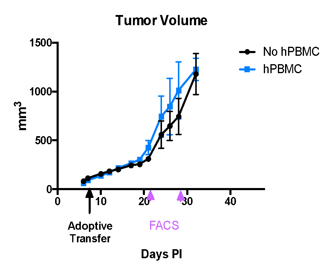
- Experience in intravenous transfer of CAR T-cells (or human PBMCs) into a mouse and in vivo / ex vivo CART therapeutic evaluations for liquid and solid tumors.
- Experience in cell line derived xenograft- in humanized mouse models and non-invasive monitoring of tumor progressions in animals.
- Detection of tumor-specific antigens by flow cytometry, in vitro or in vivo pre-screening of human PBMCs.
- Same/next day shipment of biological samples from our research facility at Morgan Hill, CA.